

The equipment behind every accurate diagnosis
Precise eye care starts with precise instruments. Our suite of advanced diagnostic technology supports everything we do, from swift, accurate refraction to the early detection of sight-threatening conditions and the fitting of our custom-designed scleral lenses.
The eye test, without the guesswork
Our Nidek RT-3100 Intelligent Refraction Unit revolutionises the traditional eye examination. Say goodbye to the tedious "1 or 2" lens clicks: this automated system offers smooth, rapid lens selection for quicker, less fatiguing and highly accurate eye examinations. Seamlessly integrated with a digital chart, it provides diverse chart options, removing the need for memorisation and catering for all ages and literacy levels.
Comparing against your old spectacles is now possible too. Lens movement is smooth and almost instantaneous, so you can genuinely appreciate the change in your prescription, if there is any at all.

Exploring the depths of eye health
Fundus photography enables non-invasive exploration of the posterior segment of the eye. Beyond assessing ocular health, it reveals insights into systemic diseases such as diabetes, multiple sclerosis and hypertension. Within the limited view through the pupil we observe the critical macula and the optic nerve, the conduit for visual information to the brain. It aids the early detection of conditions including diabetic retinopathy, glaucoma, retinitis pigmentosa and age-related macular degeneration.


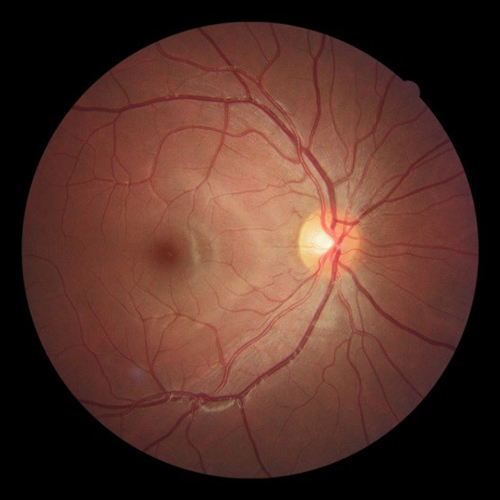


All clinical images are shown in full, uncropped. Select any image to view it at full size.
Mapping corneal precision
Our corneal topography system constructs a detailed 3D map of the cornea. It serves many roles, from assessing corneal shape to diagnosing conditions such as keratoconus. Notably, it plays a pivotal role in the design of custom contact lenses, offering a cost-effective and time-efficient approach by simulating multiple virtual fits before an actual lens is produced.
There are many cases where patients have been unable to improve their vision with spectacles, and the topographer has shown why, detecting surface abnormalities on the cornea from dry eye to ulcers. As a diagnostic tool it can also detect meibomian gland dysfunction, one of the main causes of dry eye.


Technology in the service of clinical judgement
Instruments do not fit lenses or diagnose disease; experienced clinicians do. What this equipment provides is the detail: earlier detection of sight-threatening conditions, and the precision behind every custom scleral lens and Ortho-K fit we design.
Illuminating the front of the eye
The slit-lamp biomicroscope provides high-resolution, magnified views of the anterior structures of the eye. It is instrumental in diagnosing a spectrum of conditions, including dry eye, foreign body injuries, red eye presentations and lid anomalies. It is also an indispensable tool in assessing the fit of contact lenses, ensuring optimal comfort and vision.


OCT: vision precision beyond compare
Our OCT system functions much like a medical CAT scan, delivering profiled and magnified images of the eye. In ophthalmology, OCT is indispensable for the early detection of sight-threatening diseases such as glaucoma and macular degeneration. It is also instrumental in refining the fit of our custom-designed scleral lenses, providing an intricate, close-up view of the interaction between lens and eye.


All clinical images are shown in full, uncropped. Select any image to view it at full size.
Elevate your eye care experience with us
From swift, precise refraction to the early detection of sight-threatening conditions, our advanced diagnostic instruments ensure the highest standard of care. Book your examination and experience precision like never before.
Landline & WhatsApp: 011 472 7430 · info@paulmoldovanos.co.za